
Obstetrics & Pelvic Ultrasound
In-house early viability scans, NT scan, 18-22 week anomaly TIFFA scan, 3D/4D fetal Doppler, and pelvic sonography by Dr. Manisha Kumari.

Dr. Manisha Kumari
MBBS, MS (O&G) Consultant Gynaecologist & Ultrasound Specialist- Dumduma Clinic: Near SBI, Phase-1
- IMS & SUM Hospital: K8, Kalinga Nagar
Best Obstetrics Ultrasound Scans in Bhubaneswar
High-resolution 3D/4D fetal scans, 11-13 week NT/NB screening, 18-20 week TIFFA anomaly scans, and fetal growth Doppler performed personally by Dr. Manisha Kumari in Bhubaneswar.
Dr. Manisha Kumari
MBBS, MS (O&G) Consultant O&G & Obstetric Sonologist- Dumduma Clinic: Near SBI, Phase-1
- IMS & SUM Hospital: K8, Kalinga Nagar
High-Precision Pregnancy Sonography
- Are you due for your 11-13 week NT/NB scan or 18-20 week Level-2 TIFFA anomaly scan?
- Do you need a growth Doppler scan to monitor amniotic fluid (AFI) or placental blood flow?
- Would you prefer your scans performed and explained personally by a senior obstetrician?
Obstetric ultrasound is the single most critical diagnostic window into your developing baby’s health, growth, and structural development. Dr. Manisha Kumari brings specialized sonology training and advanced 3D/4D ultrasound equipment to provide accurate, reassuring scans throughout pregnancy.
By combining maternal obstetric clinical care with direct in-house sonography at Dumduma Clinic and IMS & SUM Hospital, Dr. Manisha eliminates third-party delays and provides instant clinical answers to expectant parents.
- 1. Early Dating & Viability Scan (6 to 8 Weeks): Confirms an intrauterine gestational sac, detects embryonic heartbeat (cardiac flicker), establishes accurate gestational age (EDD), and rules out ectopic or molar pregnancy.
- 2. NT / NB Genetic Screening Scan (11 to 13.6 Weeks): Measures fetal nuchal translucency (NT thickness) and visualizes the nasal bone (NB) to screen for Down syndrome and major chromosomal/cardiac abnormalities.
- 3. TIFFA / Level-2 Anomaly Scan (18 to 20 Weeks): Meticulous organ-by-organ evaluation of fetal brain, face, palate, four-chamber heart, spine, stomach, kidneys, abdominal wall, and limbs, alongside placental localization and cervical length.
- 4. Fetal Growth & Color Doppler Scan (28 to 36 Weeks): Serial estimation of fetal weight (EFW), amniotic fluid index (AFI), and Doppler vascular resistance in umbilical and middle cerebral arteries to confirm fetal vitality.
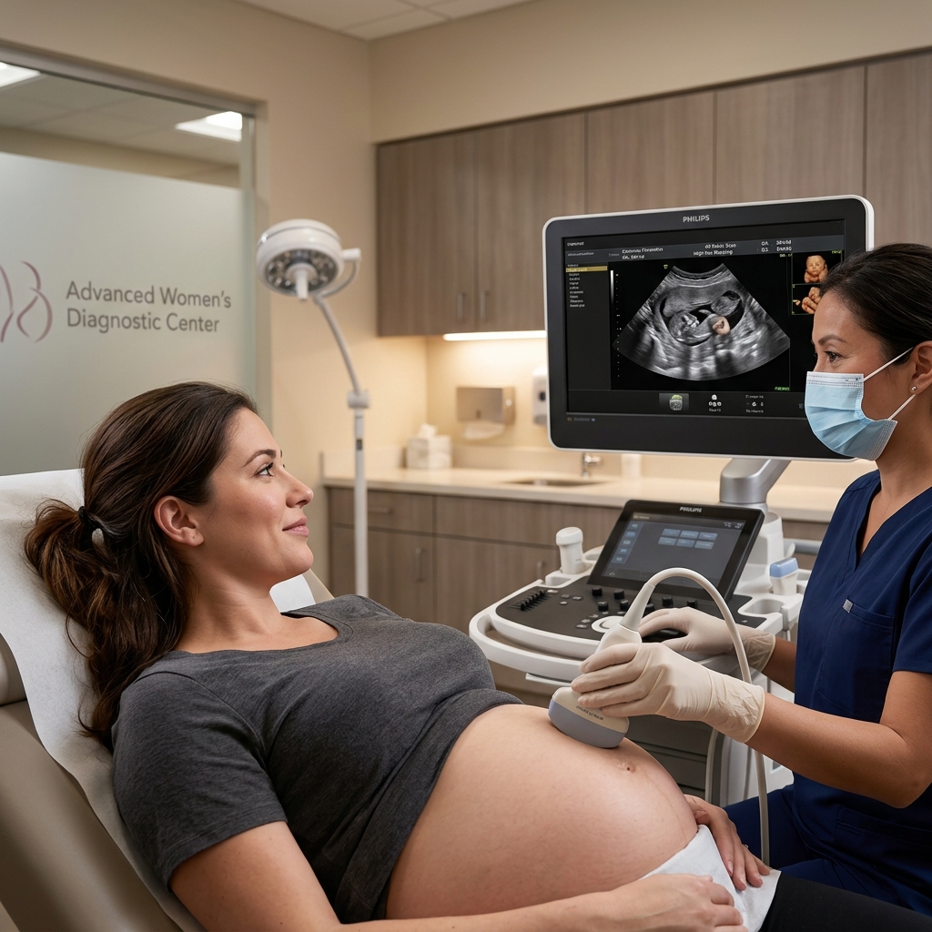
Parents can watch their baby on the high-definition monitor while Dr. Manisha explains each measurement in real time.
Why Having Your Obstetrician Perform Your Scan Matters
Doctor-Led In-House Ultrasound
- The doctor who delivers your baby performs and understands your scan.
- Immediate clinical explanation during the scan—zero anxiety or waiting.
- Immediate management adjustments if growth lag or low fluid is detected.
- Seamless continuity of care under one roof without visiting external labs.
Standalone Commercial Diagnostic Labs
- Technicians cannot prescribe medications or answer clinical questions.
- Hours or days of waiting for printed report delivery.
- Frequent unnecessary panic over normal anatomical variants.
- Requires multiple appointments and separate visits across different locations.
Affordable Transparent Pricing
All pregnancy ultrasound scans adhere strictly to the PC-PNDT Act, offering transparent and affordable rates in Bhubaneswar.
In-house sonography conducted at Dumduma Clinic and IMS & SUM Hospital, Bhubaneswar.
Why Choose Dr. Manisha Kumari for Pregnancy Ultrasound?
- Specialized Ultrasound Fellowship: Clinical training dedicated to high-precision fetal imaging and anomaly detection.
- Scans Performed Personally: No third-party technicians—Dr. Manisha performs every scan herself.
- Immediate Clinical Integration: Same-day scan reporting and immediate antenatal consultation under one roof.
- State-of-the-Art Color Doppler: Accurate assessment of placental health, uterine resistance, and fetal growth.
- Patient, Reassuring Environment: Ample time provided to answer questions and view your baby in comfort.
- Strict Ethical Compliance: Full adherence to PC-PNDT guidelines, promoting fetal health and well-being.
- Dual Practice Locations: Dumduma Clinic OPD & IMS & SUM Hospital, Bhubaneswar.

Frequently Asked Questions
Common questions regarding pregnancy ultrasound scans, safety, and preparation.
Yes, 100% safe. Diagnostic ultrasound uses high-frequency sound waves (not ionizing radiation or X-rays). Over four decades of global clinical evidence confirm that routine diagnostic ultrasound has no harmful effects on fetal growth or health.
For early scans (before 10 weeks) done abdominally, a moderately full bladder is helpful. However, for early transvaginal scans (TVS) and for all scans in the second and third trimesters (such as TIFFA and Growth Doppler), an empty bladder is preferred for optimal comfort and imaging.
A Level-1 scan is a basic check of fetal heartbeat, gestational age, and placental position. A Level-2 (TIFFA) scan is an exhaustive 20-to-30 minute examination that inspects the micro-anatomy of every major fetal organ (brain ventricles, heart chambers, facial clefts, kidneys, spine, and limbs) to rule out structural anomalies.
Yes, absolutely. We encourage partners to be present to experience the joy of seeing their baby's movements and heartbeat on the live viewing screen.